Subtyping of pancreatic cancer with 3D-organoid-models
Bösherz Lab
Our group works on characterizing and stratifying pancreatic cancer on the histomorphological an molecular level with the goal to help identify new therapeutic targets and ultimately improve the prognosis of this still aggressive and lethal disease.
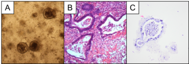
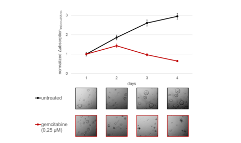
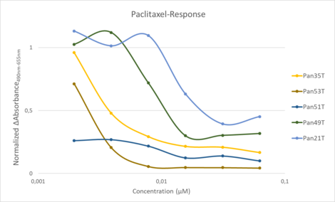
To this end we have established and now utilize primary 3D-tissue-culture-models, so called „organoids“, of pancreatic cancer specimens resected from patients in the UMG. After successful isolation and cultivation, the individual organoid-lines are admissible for molecular characterization and manipulation as well as testing for sensitivity against conventional or new therapeutics.
Members
Dr. med. Mark-Sebastian Bösherz
Jennifer Appelhans
cand. med. Antonia Johanna Faller
Collaborations
Our projects are designed and carried out in close cooperation with the AG Papantonis, AG Conradi of the Department of General, Visceral and Child Surgery and the AG Heßmann of the Department of Gastroenterology and Gastrointestinal Oncology and funded by the German Research Foundation as part of the Clinical Research Unit (DFG KFO 5002).